Introduction to Research Method in Neuropsychology
Research methods in neuropsychology are very important to gain wholesome understanding of biopsychology, which essentially comprises neuroanatomy, neurophysiology and neurochemistry. These methods are invaluable for advancing our understanding of:
- Brain-behavior relationships.
- Neural mechanisms of diseases like Alzheimer’s, Parkinson’s, or stroke.
- Effects of treatments, such as drugs or brain stimulation.
- Developmental and cognitive processes.
Know more about scope of neuropsychology
Anatomical Research methods in neuropsychology
There are three key neuropsychological research methods that provides information about anatomy i.e structure of the brain. they are – contrast x-rays, CT and MRI scanning.
Contrast x-rays
- Normal X-rays are not beneficial to get a clear picture of brain, since it has many overlapping internal structures that are NOT show different degree of absorption of x-ray radiation. Read about Working of X-rays.
- Instead, a contrast (a chemical substance) is injected into specific part of the brain, which gives it differentiating contrasting power. One contrast x-ray technique, cerebral angiography, uses the infusion of a radio- opaque dye into a cerebral artery to visualize the cerebral circulatory system during x-ray photography.
- Cerebral angiograms are most useful for localizing vascular damage, but the displacement of blood vessels from their normal position also can indicate the location of a tumor.
2. Computerized Tomography (CT)
- CT is a computer-assisted X-ray procedure that generates detailed images of internal structures, including the brain. It provides cross-sectional (horizontal) views that can be compiled into three-dimensional representations.
- CT Procedure:
- The patient lies on a table with their head positioned in the centre of a large cylindrical scanner.
- An X-ray tube emits a beam through the patient’s head. A detector on the opposite side captures the transmitted X-rays.
- The X-ray tube and detector rotate around the patient’s head at a specific level, capturing numerous images from different angles.
- The captured X-ray data is processed by a computer to create a CT scan of a horizontal section of the brain.
- The X-ray apparatus is moved to successive levels along the body axis to capture images of multiple horizontal brain sections.
- Combining scans from different levels allows for a 3D representation of the brain’s structure.
3. Magnetic Resonance Imaging (MRI)
- Magnetic Resonance Imaging (MRI) is a high-resolution brain imaging technique that constructs detailed anatomical images by measuring the electromagnetic energy emitted by hydrogen atoms in response to a powerful magnetic field.
- Principle of MRI
- A powerful magnetic field, about 25,000 times stronger than the Earth’s magnetic field, is applied to align the axes of rotation of hydrogen nuclei in the body.
- A brief radio-frequency field is introduced, tilting the aligned hydrogen nuclei.
- When the radio-frequency field is switched off, the hydrogen nuclei return to their original alignment, releasing electromagnetic energy during the process.
- The emitted electromagnetic energy is measured and processed to create detailed images of brain structures.
- MRI have very high spatial resolution, detecting structures as small as a millimeter. MRI produces superior images compared to CT, especially for soft tissue differentiation.
- It can produce 3D images, providing a comprehensive view of the brain’s anatomy.
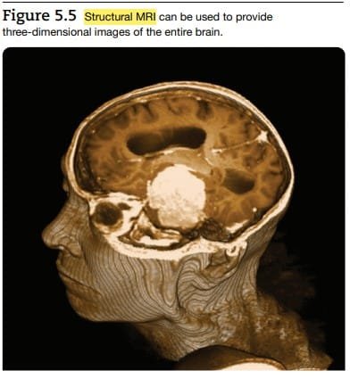
3D MRI scan. J. Pinel & S. Barnes (2018). Biopsychology. 10th ed.
Other Scanning research methods in neuropsychology
1. Positron Emission Tomography (PET):
PET was the first brain imaging technique to provide functional brain images rather than structural images. It uses radioactive fluorodeoxyglucose (FDG), which mimics glucose—the brain’s primary energy source. The process involves:
- Administered via the carotid artery, FDG is rapidly absorbed by active brain cells. However, unlike glucose, FDG is not metabolized and accumulates in active neurons or astrocytes until it breaks down (Nasrallah & Dubroff, 2013).
- PET scans map radioactivity levels in various brain areas, represented through color coding. This allows researchers to identify active brain regions during specific tasks.
Applications: PET is widely used to identify the distribution of neurotransmitters, receptors, or transporters by using radioactively labeled ligands (Camardese et al., 2014).
Limitations: PET scans provide only a colored map of brain activity and require integration with structural imaging techniques like MRI for precise localization (Wehrl et al., 2013).
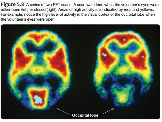
PET Scan. J. Pinel & S. Barnes (2018). Biopsychology. 10th ed.
2. Functional Magnetic Resonance Imaging (fMRI):
fMRI generates images of brain activity by measuring blood oxygenation levels (Hillman, 2014).
- Mechanism:
- Active brain regions consume more oxygenated blood than required, leading to an accumulation of oxygen.
- Oxygenated blood alters the magnetic properties of hydrogen atoms, influencing the radio-frequency signals recorded as the BOLD signal (Blood-Oxygen-Level-Dependent signal).
- Advantages of fMRI over PET:
- Simultaneously provides structural and functional information.
- Higher spatial resolution.
- Produces 3D images of brain activity.
- Limitations:
fMRI images reflect the BOLD signal rather than direct neural activity, and the complex relationship between these signals poses challenges (Hillman, 2014). Additionally, fMRI has poor temporal resolution, as it takes 2–3 seconds to measure the BOLD signal, whereas neural events like action potentials occur in milliseconds.
3. Diffusion Tensor Imaging (DTI):
DTI, an advanced form of MRI, identifies pathways where water molecules rapidly diffuse (Jbadi et al., 2015).
- Focus: By mapping water diffusion along axon tracts, DTI provides images of the brain’s major structural connections, contributing to the study of the connectome—the network of neural pathways connecting different brain regions (Calhoun et al., 2014; Lichtman, Pfister, & Shavit, 2014).
Applications: DTI is central to the Human Connectome Project, which aims to map all neural connections in the brain (Dance, 2015).
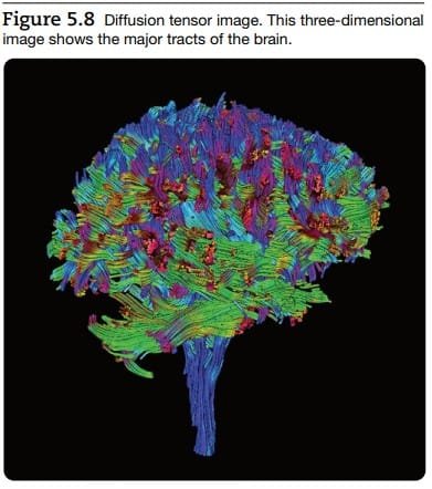
Diffusion Tensor Image (DTI). J. Pinel & S. Barnes (2018). Biopsychology. 10th ed.
4. Transcranial Magnetic Stimulation (TMS):
TMS is a non-invasive technique to assess causal relationships between brain activity and cognitive or behavioral functions.
- Mechanism: A magnetic field generated under a coil placed on the scalp either temporarily disrupts (turns off) or enhances (turns on) cortical activity (Rossini et al., 2015).
- Applications: TMS helps establish causation in areas where correlational imaging methods like fMRI or PET fall short.
5. Transcranial Direct Current Stimulation (tDCS):
tDCS applies a weak electrical current through scalp electrodes to stimulate cortical regions.
- Effects: Increases neural activity in the targeted area, enabling researchers to assess its impact on cognition and behavior (Brunoni et al., 2012).
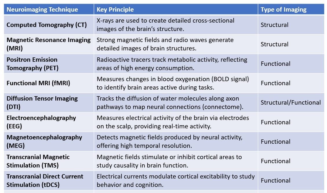
Summary : Neuroimaging techniques
Invasive Neuropsychological Research methods
Invasive techniques are the research methods that are more direct techniques, generally practice on laboratory animals which involve physically entering or manipulating the brain & nervous system. It typically require surgical procedures or interventions.
1. Stereotaxic surgery
Stereotaxic surgery is a precise technique used in biopsychology and neuroscience to position experimental devices, such as electrodes or cannulas, into specific regions of the brain. This method is foundational for experiments that require direct access to deep brain structures.
Key Components of Stereotaxic Surgery
- Stereotaxic Atlas
- Serves as a “map” of the brain, much like a geographic atlas for landmarks.
- Represents the three-dimensional structure of the brain using a series of two-dimensional frontal slices.
- Provides millimeter-precise coordinates for specific brain regions relative to a designated reference point, such as bregma in many animal atlases.
- Stereotaxic Instrument
- Comprises two main parts:
- Head Holder: Secures the subject’s head in the required position.
- Electrode Holder: Holds the experimental device, such as an electrode or cannula.
- Features a system of precision gears for movement along three axes:
- Anterior–Posterior (A–P): Front to back.
- Dorsal–Ventral (D–V): Top to bottom.
- Lateral–Medial (L–M): Side to side.
- Comprises two main parts:
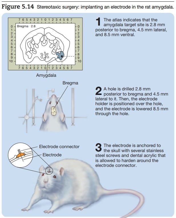
Stereotaxic surgery. J. Pinel & S. Barnes (2018). Biopsychology. 10th ed.
2. Lesion Method
Lesion methods are valuable tools for understanding brain functions by damaging, destroying, or temporarily inactivating specific brain regions and assessing the resulting behavioral effects. These methods are particularly useful for uncovering the role of specific structures in cognitive, emotional, or physiological processes.
Types of Lesions
- Aspiration Lesions
- Process: Suction is used to remove cortical tissue through a fine-tipped glass pipette.
- Advantages:
- Precise for removing surface cortical layers.
- Underlying white matter and major blood vessels can be preserved.
- Applications: Ideal for cortical regions accessible to direct visualization.
- Radio-Frequency Lesions
- Process: High-frequency current is passed through a stereotaxically positioned electrode, generating heat to destroy target tissue.
- Advantages:
- Effective for subcortical lesions.
- Size and shape of the lesion can be controlled by adjusting the current’s duration and intensity.
- Applications: Commonly used for deep brain structures.
- Knife Cuts
- Process: A small, well-placed incision is made to sever a nerve or tract without damaging adjacent tissues.
- Advantages:
- Minimal impact on surrounding structures.
- Applications: Useful for studying the role of specific neural pathways or tracts.
- Reversible Lesions
- Process: Temporarily inactivate brain regions using cooling or anesthetic injections (e.g., lidocaine).
- Advantages:
- Non-destructive.
- Allows repeated testing in both lesioned and control conditions on the same subject.
- Applications: Ideal for studies requiring reversible functional disruptions.
Key Considerations in Lesion Studies
- Interpretation Challenges (limitation): Lesion effects can be misleading due to the complexity and proximity of brain structures. Damage to neighboring areas can confound interpretations of behavioral changes. Lesions often referred to as targeting a specific structure (e.g., the amygdala) may inadvertently affect adjacent regions, complicating conclusions.
- Bilateral vs. Unilateral Lesions:
- Unilateral Lesions:
- Restricted to one hemisphere.
- Often produce milder behavioral effects, especially in non-human species.
- Bilateral Lesions:
- Involve both hemispheres.
- Typically result in more pronounced and detectable behavioral changes.
- More commonly used in experimental research.
- Unilateral Lesions:
Tools Used to Measure Neuron’s Electrical Activities
1. Oscilloscope
- The oscilloscope was a key tool in Hodgkin and Huxley’s groundbreaking experiments (1936). It functions as a highly sensitive voltmeter, capable of graphically displaying the small electrical signals generated by neurons or nerves over time.
- The vertical axis of the oscilloscope represents electrical charge in millivolts (mV), while the horizontal axis represents time in milliseconds (ms).
- The oscilloscope’s sensitivity made it possible to record the minuscule voltage changes across a neuron’s membrane.
- While modern recording systems interfaced with computers have largely replaced oscilloscopes, their role in foundational neuroscience research remains monumental.

Method of research in neuropsychology : Oscilloscope. Kolb and Whishaw (2009). Fundamentals of Human Neuropsychology
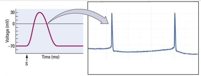
On the graph of a trace produced by an oscilloscope, S stands for stimulation. The horizontal axis measures time, and the vertical axis measures voltage. By convention, the axon voltage is represented as negative, in millivolts (mV). On the right, one trace of two action potentials from an individual neuron as displayed on a digital oscilloscope screen.
2. Microelectrodes
Microelectrodes are essential for measuring and manipulating the electrical activity of neurons. These finely-tipped electrodes are small enough to interact with individual axons and come in two main forms:
- Metal Microelectrodes: Made by etching thin wire to a fine point (approximately 1 micrometer) and insulating the rest of the wire. These electrodes can be placed directly on or inside an axon to measure electrical currents.
- Glass Microelectrodes: Constructed from thin glass tubes with tapered, fine tips (as small as 1 micrometer). When filled with a conductive salty solution, the glass microelectrode can measure voltage changes or deliver currents to neurons.
Microelectrodes offer versatile recording capabilities:
- Extracellular Recording: The tip of the electrode is placed on the axon’s surface to measure electrical currents outside the cell.
- Intracellular Recording: One electrode tip is placed inside the axon, while another remains outside, allowing the measurement of voltage differences across the neuronal membrane.
- Patch-Clamp Technique: A refined method where the tip of a glass microelectrode is suction-sealed to a small patch of the neuron’s membrane. This technique isolates and records electrical activity from a specific membrane segment.
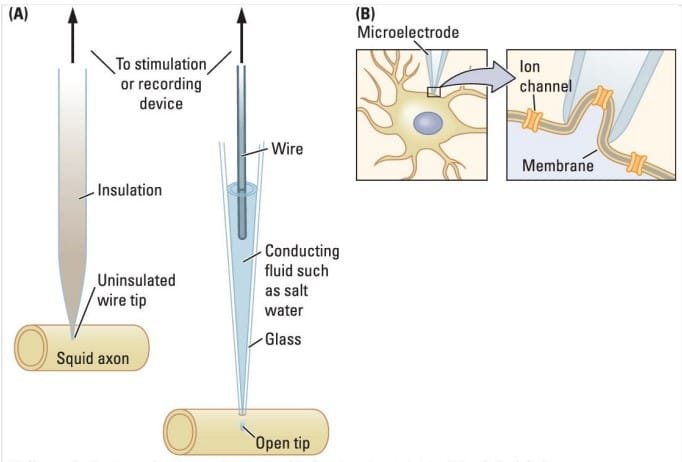
Method of research in neuropsychology : Microelectrode. Kolb and Whishaw (2009). Fundamentals of Human Neuropsychology
Applications in Hodgkin and Huxley’s Research
Using the giant axon of the squid, microelectrodes, and an oscilloscope, Hodgkin and Huxley demonstrated that the nerve impulse results from changes in ion concentrations across the neuronal membrane. Their experiments established that intracellular and extracellular ions, carrying positive and negative charges, are the basis of neuronal electrical activity.
Watch an interesting video on Giant Squid Axon
Pharmacological (Chemical) research methods to neuropsychology
Psychopharmacology employs chemical methods to study brain functions by altering neurotransmitter activity and observing behavioral consequences.
Routes of Drug Administration
Drugs are administered in psychopharmacological studies through the following methods:
- Peripheral Routes
- Oral: Fed to the subject.
- Intragastric (IG): Injected into the stomach.
- Intraperitoneal (IP): Injected into the abdominal cavity.
- Intramuscular (IM): Injected into a large muscle.
- Subcutaneous (SC): Injected beneath the skin.
- Intravenous (IV): Injected directly into a vein.
Limitation: Many drugs fail to cross the blood-brain barrier effectively.
- Central Route : Cannula Injection: Drugs are stereotaxically implanted into specific brain regions using a fine tube, bypassing the blood-brain barrier.
There three key techniques to study neuropsychology through chemical methods – (A) Selective chemical lesions, (B) Techniques measuring brain activities (C) Techniques for Locating Neurotransmitters and Receptors
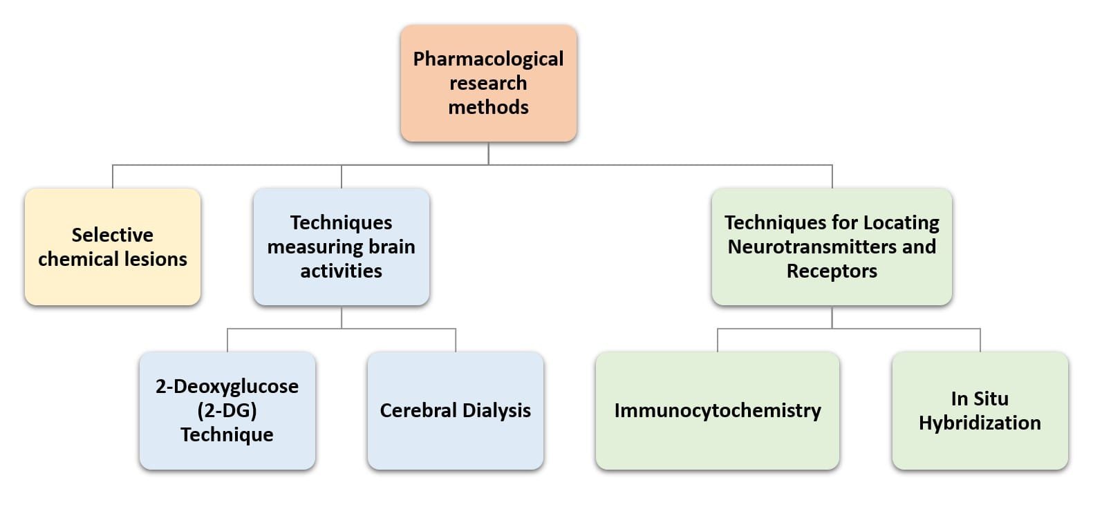
Chemical research methods in neuropsychology
A. Selective Chemical Lesions
Selective chemical lesion methods enable researchers to target specific neurons or neurotransmitter systems, offering greater precision than traditional lesion techniques.
Neurotoxin Use:
- Kainic Acid & Ibotenic Acid: Destroy cell bodies at the cannula tip while sparing axons passing through the area.
- 6-Hydroxydopamine (6-OHDA): Selectively targets neurons that release norepinephrine or dopamine without affecting neighboring cells.
Advantage: Selective chemical lesions reduce the confounding effects of damaging adjacent neurons.
B. Techniques for Measuring Chemical Activity in the Brain
- 2-Deoxyglucose (2-DG) Technique
The 2-DG method identifies brain regions with heightened activity during specific tasks by tracking glucose analog uptake. It identifies neural circuits activated during specific behaviors. It is useful in understanding functional neuroanatomy.
Procedure:
- The animal is injected with radioactive 2-deoxyglucose, a glucose analog that neurons absorb but do not metabolize.
- The subject engages in a task of interest, leading to increased neuronal activity in brain regions involved in the task.
- Post-Test Analysis:
- The animal is euthanized, and its brain is removed, sliced, and prepared for autoradiography.
- Slices are coated with photographic emulsion and stored in the dark.
- Developed slides reveal brain regions with high 2-DG absorption as black spots, which can be color-coded for detailed analysis.
2. Cerebral Dialysis
Cerebral dialysis measures extracellular neurochemical concentrations in live, behaving animals, allowing real-time monitoring of brain activity. It tracks changes in neurotransmitter levels during specific behaviors or experimental conditions. It enables longitudinal studies of neurochemical dynamics without euthanizing the subject.
Procedure:
- A fine tube with a semipermeable section is implanted in the brain region of interest.
- Neurochemicals in the extracellular fluid diffuse into the semipermeable section of the tube.
- Extracted neurochemicals are either- Stored for later analysis, or sent directly to a chromatograph for real-time chemical composition measurement.
C. Techniques for Locating Neurotransmitters and Receptors
- Immunocytochemistry
- Principle: Uses antibodies that bind to specific neuroproteins or their synthesizing enzymes.
- Procedure:
- Inject a foreign protein (antigen) to stimulate the creation of antibodies.
- Label antibodies with dyes or radioactive markers.
- Expose brain slices to labeled antibodies to identify regions containing the target neuroprotein.
- Applications:
- Locating neurotransmitters by tagging enzymes involved in their synthesis.
- Mapping neuropeptides and their receptors in the brain.
- In Situ Hybridization
- Principle: Targets mRNA sequences that direct the synthesis of specific neuroproteins.
- Procedure:
- Synthesize hybrid RNA strands complementary to the target mRNA sequence.
- Label hybrid RNA strands with dyes or radioactive markers.
- Expose brain slices to the hybrid RNA strands to identify neurons releasing the target neuroprotein.
- Applications: Mapping neurons that express specific peptides or proteins.
Polygraph
Polygraphy, commonly known as the “lie detector test,” measures autonomic nervous system (ANS) activity, such as heart rate, skin conductance, and blood pressure, to infer truthfulness during interrogations. It assumes that lying elicits heightened emotional responses, which manifest in measurable physiological changes (Ben-Shakhar, 2012).
However, polygraphy DOES NOT DIRECTLY DETECT LIES but instead captures emotional arousal, which makes its reliability limited in real-world scenarios. Emotional reactions to questions, irrespective of guilt or innocence, can confound results.
Autonomic Nervous System (ANS) Activity in Polygraphy : Polygraph tests measure physiological changes driven by the ANS. These include:
- Electrodermal Activity (EDA):
- Reflects changes in skin conductance caused by sweat gland activation during emotional arousal.
- Key Measures:
- Skin Conductance Level (SCL): Background level of skin conductivity in a given situation.
- Skin Conductance Response (SCR): Transient changes in conductivity associated with discrete emotional experiences.
- Sweating increases conductivity, especially on the hands, feet, and forehead (Green et al., 2014).
- Cardiovascular Activity:
- Heart Rate: Recorded via an electrocardiogram (ECG/EKG); emotional triggers (e.g., fear) increase heart rate.
- Blood Pressure: Measured as systolic/diastolic pressure (e.g., 130/70 mmHg is normal; >140/90 mmHg indicates hypertension).
- Blood Volume: Local changes in blood flow are associated with specific psychological events, such as sexual arousal.
3. Plethysmography: Plethysmography techniques assess changes in blood volume in specific body parts, such as fingers or genitals, which are influenced by emotional states:
- Strain Gauge Plethysmography: Measures blood flow by wrapping a gauge around the target tissue.
- Light Absorption Method: Uses light to detect blood volume changes; more blood absorbs more light.
Key Methods in Polygraphy
- Control-Question Technique (CQT):
- Compares physiological responses to target questions (e.g., “Did you steal that purse?”) with responses to control questions (e.g., “Have you ever been in jail?”).
- Lying is presumed to produce greater sympathetic activation.
- Success Rate: Approximately 80% in mock-crime studies (Ben-Shakhar, 2012).
- Guilty-Knowledge Technique (GKT):
- Detects the suspect’s reaction to details of the crime known only to the perpetrator (e.g., “Where was the purse found: in the washroom or locker?”).
- Focuses on specific crime-related knowledge rather than general emotional arousal.
- Success Rate: In controlled studies, GKT identified 88% of guilty individuals without falsely accusing the innocent (Lykken, 1959).
Conclusion
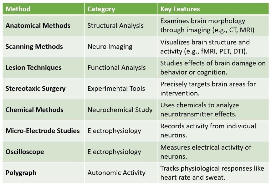
Summary of reseach methods in neuropsychology
Read more on Neuropsychology
References
- Galloti, K. M. (2004). Cognitive psychology in and out of the laboratory. USA: Thomson Wadsworth.
- Kalat, J. W. (2019). Biological psychology. Cengage.
- Kolb, B., & Whishaw, I. Q. (2019). An introduction to brain and behavior.
- Matlin, M. (1994). Cognition. Bangalore: Harcourt Brace Pub.
- Pinel, J. (2023). Biopsychology 10th Edition. Pearson.
- Schwiening C. J. (2012). A brief historical perspective: Hodgkin and Huxley. The Journal of physiology, 590(11), 2571–2575. https://doi.org/10.1113/jphysiol.2012.230458
Subscribe to Careershodh
Get the latest updates and insights.
Join 16,603 other subscribers!
Niwlikar, B. A. (2025, January 16). 8 Important Research Methods in neuropsychology : Different ways used to study Neuropsychology. Careershodh. https://www.careershodh.com/research-methods-in-neuropsychology/
