Introduction to Neural Conduction
Neurons, the fundamental units of the nervous system, are specialized cells responsible for neural conduction i.e transmitting information throughout the body. Their primary function is to generate, receive and send electrochemical signals to other neurons, muscles, or glands, ensuring coordination and communication within the body. This signaling process is achieved through the generation and conduction of electrical charges, commonly referred to as nerve impulses.
A nerve impulse are the series of electrical signals that is generated in the neurons (nerve cells) in a response to stimulus. They are also called as Action Potential which are the basic information units which relay messages along the axon over long distances (Cotman & McGaugh, 1980).
Action Potential : remarkable mechanism of neural conduction
Action potential conducts the information encoded through sensory stimuli to brain and then to targeted area of the body. The mechanism of action potential helps to in this long distance communication without any loss. The action potential allows neurons to communicate at incredible speeds, almost around 0.1 meters per second to 100 meters per second (Feher, 2017).
Role of Electric Polarization in Neural Conduction
Electrical polarization is a difference in electrical charge between two locations (Kalat, ). Here, electric charges are produced by movement of ions inside and outside of the neuron. The positively charged Na+ & K+ ions and negatively charged Cl‾ keep continuously moving in and out. Negatively charged protein molecules are also present in the cytoplasm which do not move but contribute to polarization in neuron.
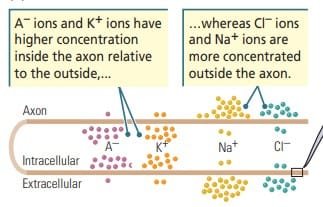
Ion distribution in neuron
3 factors influence the movement of ions
(1) Concentration gradient : The difference in distribution of ions across the membrane. Ions naturally move from areas of high concentration to low concentration (down their concentration gradient) through diffusion.
(2) Voltage gradient : The difference in electrical charge across the membrane creates a voltage gradient. Ions are attracted to regions of opposite charge (e.g., positively charged ions like Na⁺ are attracted to the negatively charged inside of the cell).
(3) Semi-permeability of cell membrane : The cell membrane’s selective permeability, determined by ion channels and transporters, controls which ions can pass through. Certain ions (e.g., K⁺) pass through more easily than others (e.g., Na⁺ or Cl⁻), influencing the membrane potential.
Recording the Membrane Potential
Hodgkin and Huxley (1936) determined how neurons send information, laying the foundation for our understanding of the electrical activity of neurons. They discovered that differences in the concentration of ions on the two sides of a cell membrane create an electrical charge across the membrane. They also revealed that this electrical charge can travel along the surface of the membrane, explaining how neurons propagate signals efficient!
To know How the Neural Conduction is studied by Read Research Methods in neuropsychology
- Electrode Placement: One electrode is placed inside the neuron (intracellular electrode). The other electrode is placed outside the neuron in the extracellular fluid.
- Intracellular Electrode (Microelectrode): The intracellular electrode must have an extremely fine tip (less than one-thousandth of a millimeter in diameter) to pierce the neuronal membrane without causing significant damage. These fine-tipped electrodes are called microelectrodes, and their precision is critical for accurate recordings.
- Extracellular Electrode: The size of this electrode is less critical compared to the intracellular one, as it does not need to penetrate the membrane.
This setup measures the voltage difference between the inside and outside of the neuron, which represents the membrane potential.
Types of Neural Potentials
There are three types of Neural potentials :
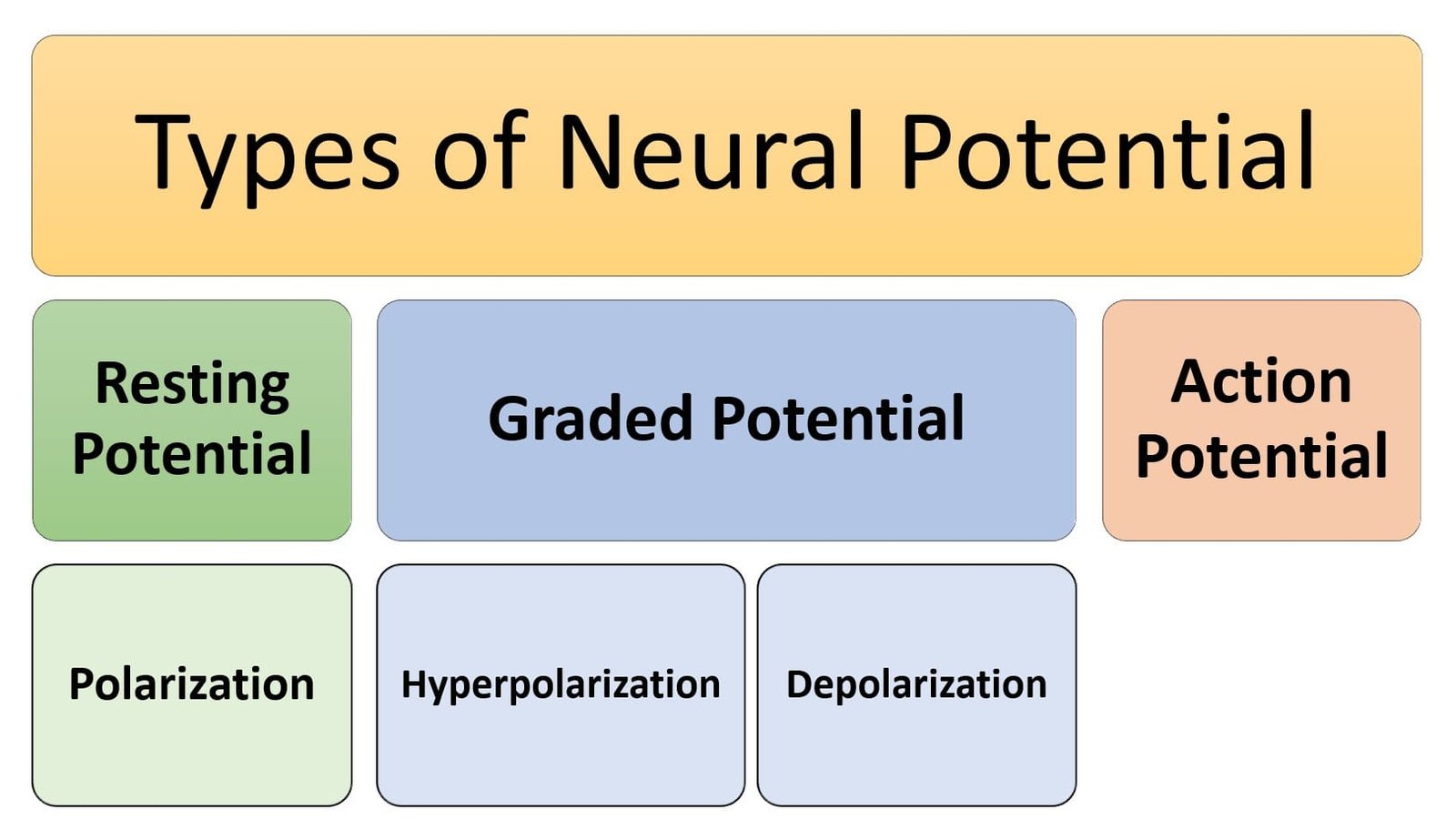
Types of neural potential
1. Resting Potential
When the tip of the intracellular microelectrode is inserted into a neuron, the voltage difference between the inside (cytoplasm) of the neuron and the extracellular fluid is measured as about −70 mV. This indicates that the inside of the neuron is 70 mV more negative than the outside. This steady, stable voltage difference across the membrane of a neuron when it is NOT actively transmitting a signal is known as Resting potential.

Resting potential
The resting potential is maintained due to the unequal distribution of ions (e.g., more sodium (Na⁺) outside the cell and more potassium (K⁺) inside the cell), contributing to the membrane’s charge. The outside of the membrane is positive, while the inside is negative, creating a −70 mV difference. In its resting state, the neuron is considered polarized, meaning there is a charge difference across its membrane.
The resting potential represents stored potential energy. Just like money in a bank account can be spent in the future, the stored charge in the membrane can be used to generate an action potential when certain conditions change.
Contributors to Resting Potential
The resting potential of a neuron is influenced by four key types of charged particles that are unequally distributed across the membrane:
- Sodium ions (Na+) : They can reduce the membrane’s resting potential by diffusing into the cell. However, the sodium channels in the membrane are mostly closed, preventing significant sodium influx. The sodium-potassium pump is crucial in maintaining the high concentration of Na⁺ ions outside the cell.
2. Potassium ions (K+) : They accumulate inside the neuron to balance the negative charge of protein anionst. Yet, they do not fully neutralize the negative charge of protein anions, contributing to the overall negative resting potential. The concentration of K⁺ ions inside the cell is about 20 times higher than outside. Due to the concentration gradient, some K⁺ ions move out of the cell, making the inside of the membrane negatively charged relative to the outside.
3. Large protein anions (A⁻) : They are synthesized inside the cell and cannot pass through the membrane. They contribute to the negative charge inside the cell, playing a key role in the transmembrane voltage gradient.
4. Chloride ions (Cl⁻) : Chloride ions move freely in and out of the cell through chloride channels in the membrane. At equilibrium, the concentration gradient for Cl⁻ ions is balanced by the voltage gradient, contributing little to the overall resting potential.
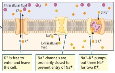
Ions contributing to Resting Potential
Sodium-Potassium Pump
The sodium-potassium pump is essential for maintaining the resting membrane potential and creating the proper ion gradient. Sodium ions are present in higher concentration outside the neuron, while potassium ions are more concentrated inside the cell. The pump helps maintain this imbalance. The imbalance is essential for the neuron to be in a “ready” state to fire an action potential. The polarized state of neuron is vital for the generation of electrical signals. Without this active pumping, the ions would gradually diffuse to equalize concentrations, disrupting the cell’s resting potential.
The pump moves three Na⁺ ions out of the neuron and two K⁺ ions into the neuron for every cycle, against their concentration gradients.
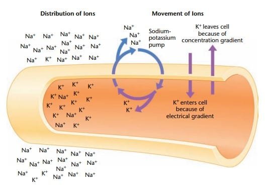
Sodium Potassium Pump
This continuous pumping action prevents Na⁺ from leaking into the cell and reduces the likelihood of the resting potential being diminished.
2. Graded Potential
A graded potential is a localized change in membrane voltage that occurs when an axon receives some form of stimulation. This change is temporary and decreases in strength as it spreads away from the site of initiation. Graded potentials are classified based on the direction of the change in membrane potential: hyperpolarization (more negative) or depolarization (less negative).
- Hyperpolarization: The membrane potential becomes more negative (e.g., -70 mV to -73 mV). This typically occurs due to an efflux of K⁺ ions or an influx of Cl⁻ ions.
- Depolarization: The membrane potential becomes less negative (e.g., -70 mV to -65 mV). This usually results from an influx of Na⁺ ions.

Types of Graded Potential
Causes of Graded Potentials
- Electrical Stimulation: A change in the flow of ions across the membrane, through ion channels or gates, causes the membrane voltage to change. The change can be brief and localized to the site of stimulation.
- Neurotransmitter Action: Neurotransmitters bind to postsynaptic receptors and either depolarize or hyperpolarize the postsynaptic membrane.
- Excitatory postsynaptic potentials (EPSPs): These are depolarizing graded potentials that increase the likelihood of a neuron firing an action potential.
- Inhibitory postsynaptic potentials (IPSPs): These are hyperpolarizing graded potentials that reduce the likelihood of a neuron firing an action potential.

Postsynaptic potentials
Characteristics of Graded Potentials:
- The amplitude of a graded potential is directly proportional to the intensity of the stimulus. A stronger stimulus causes a larger graded potential.
- As graded potentials travel away from the site of initiation, they decrease in strength. They typically travel only a short distance before they decay, meaning they don’t propagate far down the axon.
- Graded potentials can summate (combine) at the axon’s initial segment. If enough depolarization occurs and the membrane potential reaches a threshold (around -65 mV), it triggers an action potential.
- Graded potentials are integrative in that they provide the necessary input to determine whether an action potential will be initiated.
3. Action Potential
An action potential is a rapid, large change in membrane voltage that propagates along an axon. It involves a brief reversal of polarity across the membrane, which lasts around 1 millisecond. This rapid shift from negative to positive and back to negative is what allows neurons to transmit electrical signals over long distances.
All-or-none law: The amplitude and velocity of an action potential are independent of the intensity of the stimulus that initiated it. By analogy, imagine flushing a toilet: You have to make a press of at least a certain strength (the threshold), but pressing even harder does not make the toilet flush any faster or more vigorously.
Phases of Action Potential
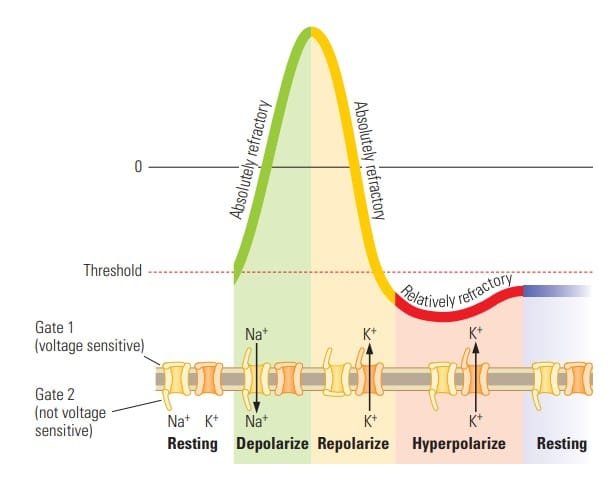
- Resting Potential: The axon starts at its resting potential of -70 mV. Ion channels are closed, and ions cannot flow through the membrane.
- Threshold and Depolarization: When the membrane reaches a threshold potential (about -50 mV), voltage-gated sodium channels open, allowing Na⁺ ions to rush into the cell. This influx causes the inside of the axon to become more positive, leading to depolarization. The membrane potential rises quickly, reaching about +30 mV.
- Repolarization: After depolarization, the sodium channels close, and voltage-gated potassium channels open. K⁺ ions leave the cell, returning the membrane potential back to negative. This phase is called repolarization.
- Hyperpolarization: As potassium continues to leave the cell, the membrane potential overshoots the resting potential, becoming slightly more negative than the resting state, resulting in hyperpolarization (around -80 mV).
- Return to Resting Potential: The potassium channels slowly close, and the membrane potential gradually returns to its resting state of -70 mV.
Phases of Action Potential
A. Generation Action Potential
- The action potential is driven by voltage-sensitive ion channels for sodium (Na⁺) and potassium (K⁺). These channels are closed at resting potential but open when the membrane reaches the threshold voltage.
- Sodium channels open more quickly than potassium channels, leading to rapid depolarization.
- Potassium channels open more slowly, leading to repolarization and hyperpolarization.
- Sodium influx (depolarization) and potassium efflux (repolarization) are what drive the action potential.
Refractory Periods
The refractory period refers to times during the action potential when the neuron cannot respond to a new stimulus:
- Absolute Refractory Period: This occurs during depolarization and repolarization phases when the sodium channels are inactivated. No new action potential can occur, regardless of the stimulus strength.
- Relative Refractory Period: After the action potential, during hyperpolarization, the membrane can respond to a new stimulus, but only if the stimulus is stronger than usual.
The refractory periods limit the frequency of action potentials, preventing backward propagation and ensuring that signals travel in one direction along the axon.
B. Conduction of Action Potential
Axonal Conduction Process
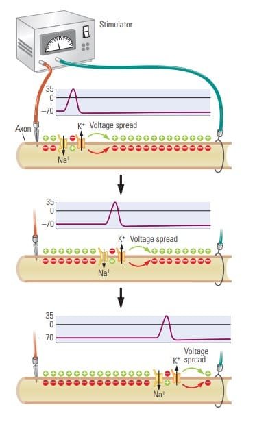
Action potentials begin at the axon hillock and propagate down the axon. This is achieved through the regeneration of the action potential along the membrane of the axon.
When an action potential is initiated, sodium (Na+) ions flow into the axon, temporarily making that region positively charged relative to the neighboring axonal areas. This positive charge flows passively along the axon and causes neighboring areas of the axon membrane to reach their threshold potential, thereby triggering new action potentials in a wave-like fashion.
Conduction of Action Potential
As the action potential travels down the axon, each segment of the membrane regenerates the action potential, ensuring the signal does not lose strength over distance. This non-decremental property contrasts with other forms of neural signaling, like EPSPs or IPSPs, which decay as they travel.
Saltatory Conduction
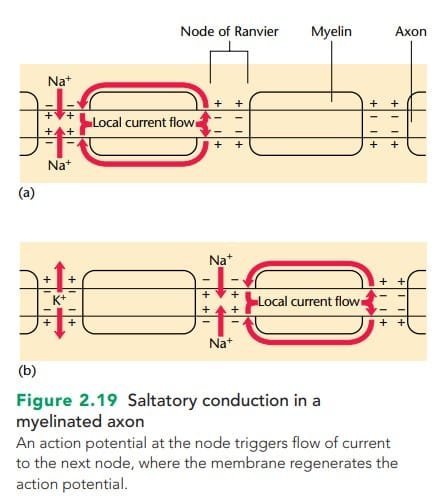
In myelinated axons, ions can only pass through the membrane at the nodes of Ranvier. Once an action potential is generated at a node, it passes passively to the next node, where it triggers another full action potential. This process is faster than the continuous conduction in non-myelinated axons and is referred to as saltatory conduction. The signal “jumps” from one node to the next, greatly increasing conduction speed.
Conduction in Non-Myelinated Axons
In non-myelinated axons, conduction is slower because the action potential travels more gradually through the axonal membrane, without the aid of saltatory conduction. The signal in these axons decays over time, thus limiting the distance the action potential can travel efficiently.
Conduction Without Axons
Not all neurons rely on axonal conduction to transmit action potentials. Some neurons, especially interneurons in the brain, do not have axons or have very short ones. These neurons typically rely on passive conduction (where the signal decays over time) rather than active conduction, which is characteristic of axons.
Using Understanding of Neural Conduction
1. Neurotoxic Substances and Their Effects:
- Scorpion Venom: This venom interferes with neural conduction by keeping sodium channels open and potassium channels closed. This causes prolonged depolarization and the dangerous accumulation of sodium ions, leading to neural dysfunction (Pappone & Cahalan, 1987; Strichartz et al., 1987).
- Tetrodotoxin: Found in puffer fish, this toxin blocks sodium channels, thereby preventing depolarization and normal action potentials. This can halt neuronal activity, which can be fatal if it disrupts critical neural functions like respiration.
- Tetraethylammonium (TEA): This chemical blocks potassium channels, preventing hyperpolarization and disrupting the normal reset of the neuronal membrane after an action potential.
2. Medical Interventions:
- Local Anesthetics (e.g., Novocain and Xylocaine): These drugs attach to sodium channels and prevent the entry of sodium ions. This blocks the generation of action potentials, particularly in sensory neurons carrying pain signals, effectively numbing the area (Ragsdale et al., 1994).
- Neurodegenerative Diseases:
- Multiple Sclerosis (MS): This condition damages the myelin sheath that insulates axons, leading to slowed or distorted neural conduction. The disease illustrates the importance of myelin in maintaining the speed and fidelity of neural communication.
Conclusion
Neural conduction refers to the transmission of nerve impulses within neurons and across synapses, essential for communication in the nervous system. It begins with the resting potential, where neurons maintain a stable, negative internal charge. Graded potentials are small, localized changes in membrane potential due to stimuli, while action potentials are rapid, all-or-nothing depolarizations that propagate along axons. Action potentials are generated when threshold levels are met, involving ion exchange through voltage-gated channels. Understanding neural conduction aids psychology by elucidating mechanisms behind perception, learning, emotions, and disorders like epilepsy or depression, bridging biological processes with behavior and mental health.
Read more about Neuropsychology
References
- Cotman, C. W., & McGaugh, J. L. (1980). Basic principles of neuronal signaling. In Behavioral Neuroscience (pp. 111–150). https://doi.org/10.1016/B978-0-12-191650-3.50009-X
- Feher, J. (2017). Propagation of the action potential. In Elsevier eBooks (pp. 280–288). https://doi.org/10.1016/b978-0-12-800883-6.00025-2
- HODGKIN, A., HUXLEY, (1939). Action Potentials Recorded from Inside a Nerve Fibre. Nature 144, 710–711. https://doi.org/10.1038/144710a0
- Kalat, J. W. (2019). Biological psychology. Cengage.
- Kolb, B., & Whishaw, I. Q. (2019). An introduction to brain and behavior.
- Pinel, J. (2023). Biopsychology 10th Edition. Pearson.
Subscribe to Careershodh
Get the latest updates and insights.
Join 16,594 other subscribers!
Niwlikar, B. A. (2025, January 19). Neural Conduction : Generation and Transmission of Action Potential. Careershodh. https://www.careershodh.com/neural-conduction-action-potential/
