
Introduction to Central Nervous System (CNS)
Anatomically, there are two types of nervous systems : Central Nervous System (CNS) and Peripheral Nervous Systems (PNS). The CNS consists of brain and spinal cord, where PNS is the division of nervous system that is located outside of the skull and spine, which include all the extending nerves reaching to every part of the body. Most of the nerves of PNS project from spinal cord. Only exception to this is 12 pairs of nerves, known as cranial nerves. They originates from the brain. Hence, Brain and spinal cord are very essential structure.
Read more about structure and types of Neurons
Support and Protection of Central Nervous System
The central nervous system (CNS) is well-protected within the body, safeguarded by bony structures. The brain is encased within the skull, a rigid and thick bone that provides critical protection against physical trauma. Similarly, the spinal cord is shielded by a series of interlocking vertebrae that form the spinal column. These bony encasements ensure the CNS is structurally supported and protected from external injuries.
Meninges
CNS is further shielded by three protective membranes known as the meninges.
- Dura Mater: The outermost layer, aptly named “tough mother,” is a robust and durable membrane that provides primary protection.
- Arachnoid Membrane: Beneath the dura mater lies this fine, spider-web-like layer. It encases the subarachnoid space, which contains large blood vessels and cerebrospinal fluid (CSF)—a vital component that cushions and nourishes the CNS.
- Pia Mater: The innermost layer, or “pious mother,” is a delicate membrane that closely adheres to the surface of the brain and spinal cord, providing additional support and nourishment.

Meninges.
Cerebrospinal fluid & Ventricles
The cerebrospinal fluid (CSF) plays a critical role in protecting and supporting the central nervous system (CNS). It fills the subarachnoid space, the central canal of the spinal cord, and the cerebral ventricles in the brain. These regions are interconnected, forming a unified reservoir that cushions and nourishes the brain and spinal cord.
Structure and Circulation
- Central Canal: A narrow channel running the length of the spinal cord.
- Cerebral Ventricles: Four large chambers in the brain:
- Lateral ventricles (2)
- Third ventricle (1)
- Fourth ventricle (1)
- Subarachnoid Space: Encases the brain and spinal cord, housing large blood vessels and CSF.
CSF circulates through these areas, produced mainly by the choroid plexuses—networks of capillaries extending from the pia mater into the ventricles. Excess CSF is absorbed from the subarachnoid space into dural sinuses, large blood-filled spaces within the dura mater, which drain into the jugular veins.
Functions of CSF
- Cushioning: CSF acts as a shock absorber, protecting the brain from impacts. Patients with reduced CSF often experience severe headaches and stabbing pains with sudden movements.
- Support: The fluid reduces the brain’s effective weight, minimizing pressure on its structures.
- Nutrient Exchange: Facilitates the transport of nutrients and waste between the brain and bloodstream.
Disorders of CSF Circulation
Hydrocephalus (“water head”) arises when CSF flow is blocked, often by a tumor obstructing narrow channels like the cerebral aqueduct (connecting the third and fourth ventricles). This blockage leads to fluid buildup, causing the ventricles and brain to expand.
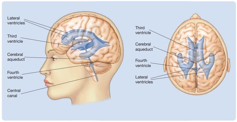
Cerebral ventricles. J. Pinel & S. Barnes (2018). Biopsychology. 10th ed.
Blood Brain Barrier
The blood-brain barrier (BBB) is a crucial defense mechanism protecting the brain from harmful substances in the bloodstream.
Structure and Function
- Unlike the loosely packed cells in blood vessels throughout the body, the blood vessels in the brain are composed of tightly packed cells. This arrangement forms a barrier that restricts the entry of large molecules, such as proteins, and many toxins.
- Essential molecules like glucose, critical for brain function, bypass the barrier through active transport mechanisms, ensuring the brain’s nutritional and functional needs are met.
Selective Permeability : The blood-brain barrier selectively permits the passage of certain substances:
- Therapeutic and recreational drugs affect brain activity depending on how easily they penetrate the BBB.
- Specific brain regions have less restrictive barriers, allowing particular molecules to pass for specialized functions.
Clinical Significance
The integrity of the blood-brain barrier is essential for neurological health. Disruptions to the BBB are linked to various central nervous system (CNS) disorders, such as neurodegenerative diseases and infections. Impaired BBB function can allow harmful substances to enter the brain, exacerbating these conditions (Bentivoglio & Kristensson, 2014).
Anatomy of Central Nervous System
1. Spinal Cord
The spinal cord is a highly organized neural structure critical for relaying sensory and motor information, coordinating reflexes, and enabling movement. Its design shares a fundamental blueprint with simpler organisms like the earthworm while showcasing increased complexity in mammals.
Cross-Section Anatomy
- Gray Matter: The inner, H-shaped region consists of neural cell bodies and unmyelinated interneurons.
- Dorsal Horns: Process sensory (afferent) inputs.
- Ventral Horns: Issue motor (efferent) outputs.
- White Matter: Surrounds gray matter and comprises myelinated axons organized into tracts.
- Dorsal Tracts/ roots: Primarily sensory, transmitting afferent signals to the brain.
- Ventral Tracts: Primarily motor, transmitting efferent signals to muscles and organs.
Bell-Magendie Law: Demonstrated that the dorsal roots are sensory, and ventral roots are motor, revolutionizing neurology by distinguishing sensory and motor impairments.
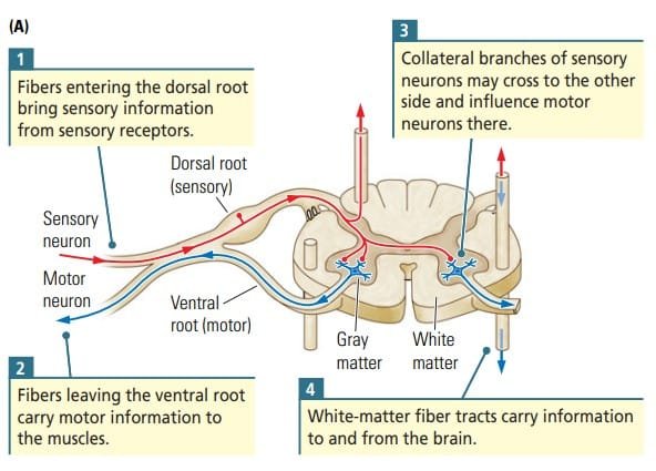
Cross section of Spinal cord. Kolb and Whishaw (2009). Fundamentals of Human Neuropsychology
Segmental Organization
- Dermatomes and Spinal Nerves
- The spinal cord comprises 31 pairs of spinal nerves, categorized by regions:
- Cervical (8): Control forelimbs and upper body.
- Thoracic (12): Govern trunk movements.
- Lumbar (5): Manage hindlimb actions.
- Sacral (5): Associated with pelvic and lower body functions.
- Dermatomes represent segments of the body innervated by specific spinal nerves. These are distorted in humans due to upright posture but maintain correspondence with spinal segments.
- The spinal cord comprises 31 pairs of spinal nerves, categorized by regions:
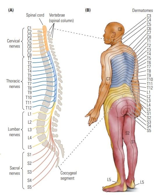
Spinal cord and spinal nerve structure. J. Pinel & S. Barnes (2018). Biopsychology. 10th ed.
Reflexes and Neural Integration
The spinal cord organizes reflexive actions, movements elicited by sensory stimulation, even in isolation from the brain.
- Flexion Reflex: Protective withdrawal of a limb in response to painful stimuli.
- Extension Reflex: Stabilization of a limb to maintain contact with stimuli, such as maintaining balance.
Sherrington’s research demonstrated that spinal cord activity could independently produce coordinated movements, such as walking, through local circuits.
5 Divisions of Brain
The The brain is an organ of nervous tissue responsible for responses, sensation, movement, emotions, communication, thought processing, and memory (Thau et. al, 2022). It is classified into five major divisions, reflecting its development from the embryonic neural tube and its functional organization. These divisions are essential for understanding brain anatomy and functions, just as knowing the neighborhoods of a city helps navigate it.

Early Development of brain. J. Pinel & S. Barnes (2018). Biopsychology. 10th ed.
Developmental Background
- During early embryonic development, the neural tube forms three primary swellings:
- Forebrain
- Midbrain
- Hindbrain
- Before birth, these three swellings further divide, forming five divisions:
- Telencephalon (anterior forebrain)
- Diencephalon (posterior forebrain)
- Mesencephalon (midbrain)
- Metencephalon (anterior hindbrain)
- Myelencephalon (posterior hindbrain, also called the medulla).
These divisions give rise to the adult brain structures.
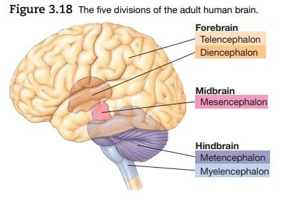
Five division of brain. J. Pinel & S. Barnes (2018). Biopsychology. 10th ed.
Brain Stem
The brain stem consists of the four divisions below the telencephalon (diencephalon, mesencephalon = midbrain, metencephalon, and myelencephalon = hindbrain). It serves as the structural base for the cerebral hemispheres. The brainstem begins where the spinal cord enters the skull and extends upward to the lower areas of the forebrain.
The brainstem facilitates more complex movements than the spinal cord but follows a similar functional layout, with the dorsal region responsible for sensory functions and the ventral region responsible for motor functions. A unique feature of the brainstem is its cranial-nerve nuclei, which converge and send axons to the head muscles. The brainstem also serves as a transit hub for fibers connecting the spinal cord and the forebrain, regulating various complex functions in its different regions.
🧠Hindbrain
1. Myelencephalon (Medulla)
- Location: Most posterior division of the brain.
- Structure:
- Composed largely of tracts transmitting signals between the brain and body.
- Contains the reticular formation: A network of about 100 tiny nuclei extending through the brainstem.
- Functions:
- Involved in basic life-sustaining functions such as cardiac, respiratory, and circulatory reflexes.
- Plays roles in arousal, sleep, attention, movement, and muscle tone.
- Misleadingly referred to as the “reticular activating system” due to its diverse functions.
2. Metencephalon
- Location: Between the myelencephalon and mesencephalon.
- Structures:
- Pons: A bulge on the ventral surface of the brainstem that relays signals between the brain and the spinal cord.
- Cerebellum (“little brain”): Located dorsally, featuring a convoluted structure.
- Functions:
- Pons: Houses ascending/descending tracts and part of the reticular formation.
- Cerebellum:
- Essential for precise movement control and adaptation to changing conditions.
- Handles equilibrium, postural reflexes
- Also involved in cognitive functions like decision-making and language.
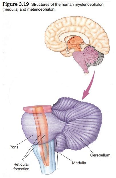
Hindbrain. J. Pinel & S. Barnes (2018). Biopsychology. 10th ed.
🧠Midbrain
3. Mesencephalon (Midbrain)
- Location: Superior to the metencephalon.
- Divisions:
- Tectum:
- Location: Dorsal surface.
- Structures: Two pairs of colliculi (bumps):
- Inferior colliculi: Auditory processing.
- Superior colliculi: Visual-motor functions (e.g., orienting toward stimuli).
- Tegmentum:
- Location: Ventral to the tectum.
- Structures:
- Reticular formation and tracts of passage.
- Periaqueductal gray: Surrounds the cerebral aqueduct, mediates opioid analgesic effects.
- Substantia nigra and red nucleus: Important for sensorimotor functions.
- Tectum:

Midbrain. J. Pinel & S. Barnes (2018). Biopsychology. 10th ed.
🧠 Forebrain
4. Diencephalon
- Location: Anterior to the mesencephalon.
- Structures:
- Thalamus:
- Description: Large, two-lobed structure at the top of the brainstem.
- Functions: Acts as a sensory relay center, processing and transmitting sensory signals to appropriate cortical areas.
- Example: Lateral geniculate nucleus (visual signals), medial geniculate nucleus (auditory signals), and ventral posterior nucleus (somatosensory signals).
- Receives feedback from cortical areas, allowing dynamic interaction.
- Hypothalamus:
- Description: Located below the thalamus.
- Functions:
- Regulates motivated behaviors such as eating, sleeping, and sexual activity.
- Controls hormone release through the pituitary gland.
- Notable structures:
- Optic chiasm: Where optic nerves partially decussate.
- Mammillary bodies: Nuclei involved in memory and learning.
- Thalamus:
5. Telencephalon
- Location: Most anterior and largest brain division.
- Structure:
- Includes the cerebral hemispheres, containing the cortex, basal ganglia, and limbic system.
- Functions:
- Basal Ganglia: Subcortical nuclei involved in movement and simple learning. Includes structures like the caudate nucleus, putamen, and globus pallidus. Disorders of the basal ganglia, such as Huntington’s chorea, Parkinson’s disease, and Tourette’s syndrome, illustrate its role in movement control and coordination.
- Limbic System: Manages emotions, motivation, and memory.
- Cerebral Cortex: Responsible for higher cognitive functions like perception, planning, and reasoning.
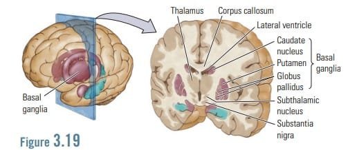
Forebrain. Kolb and Whishaw (2009). Fundamentals of Human Neuropsychology
Conclusion
The central nervous system (CNS) is a highly organized and dynamic network responsible for integrating sensory input, processing information, and coordinating bodily functions and behaviors. Structurally, it consists of the brain and spinal cord, each contributing unique yet interdependent roles. The brain, with its divisions like the forebrain, midbrain, and hindbrain, facilitates higher-order cognitive functions, emotional regulation, memory, and perception. The spinal cord serves as the conduit for neural signals between the brain and the rest of the body, enabling reflexive and voluntary movements.
Learn more about Neuropsychology
References
- Kalat, J. W. (2019). Biological psychology. Cengage.
- Kolb, B., & Whishaw, I. Q. (2019). An introduction to brain and behavior.
- Thau L, Reddy V, Singh P. Anatomy, Central Nervous System. [Updated 2022 Oct 10]. In: StatPearls [Internet]. Treasure Island (FL): StatPearls Publishing; 2024 Jan-. Available from: https://www.ncbi.nlm.nih.gov/books/NBK542179/
- Pinel, J. (2023). Biopsychology 10th Edition. Pearson.
Subscribe to Careershodh
Get the latest updates and insights.
Join 13,984 other subscribers!
Niwlikar, B. A. (2025, January 22). Central Nervous System (CNS) : Brain & Spinal Cord. Careershodh. https://www.careershodh.com/central-nervous-system-cns/